Exploring the profound symbiotic relationship between AMF and plants — with a focus on potato cultivation, soil health, and sustainable agriculture in Nepal.
✍️ Anup Pudasaini📅 January 2025🔬 Scholarly Article🌱 AMF & Phosphorus Research
80%
of all terrestrial plant species form symbiosis with AMF
400M+
years — the ancient evolutionary age of this symbiosis
50×
increase in root absorptive surface area from AMF hyphae
Introduction
The Vital Role of Plants and Plant-Fungi Symbiosis
Plants form the foundation of terrestrial ecosystems, providing food, oxygen, and resources essential for human survival and the balance of natural environments. However, their growth often faces challenges due to limited nutrient availability — especially of phosphorus (P), nitrogen (N), and other essential minerals. Efficient nutrient uptake is critical not only for maximizing crop yields but also for ensuring long-term soil fertility and sustainability.
To overcome these challenges, many plants engage in a mutualistic partnership with Arbuscular Mycorrhizal Fungi (AMF). These fungi colonize plant roots and extend their hyphae deep into the soil, increasing the effective surface area for nutrient and water absorption. In return, the plant provides the fungi with carbohydrates derived from photosynthesis.
Beyond nutrient exchange, AMF also contribute to improved soil structure, greater resistance to drought, and reduced vulnerability to soil-borne diseases. Their role extends beyond individual plants — they form vast underground networks connecting multiple plant species, facilitating nutrient sharing and supporting ecosystem stability.
🌱 Key Facts on Plant Nutrition & Sustainability
→Phosphorus plays a pivotal role in energy transfer, metabolic processes, and root development in nearly all plants.
→Excessive reliance on chemical fertilizers has adverse effects on soil health and long-term agricultural sustainability.
→AMF colonizes over 80% of terrestrial plants, significantly improving nutrient cycling and offering a natural biofertilizer alternative.
Biology
Understanding AMF: The Invisible Network
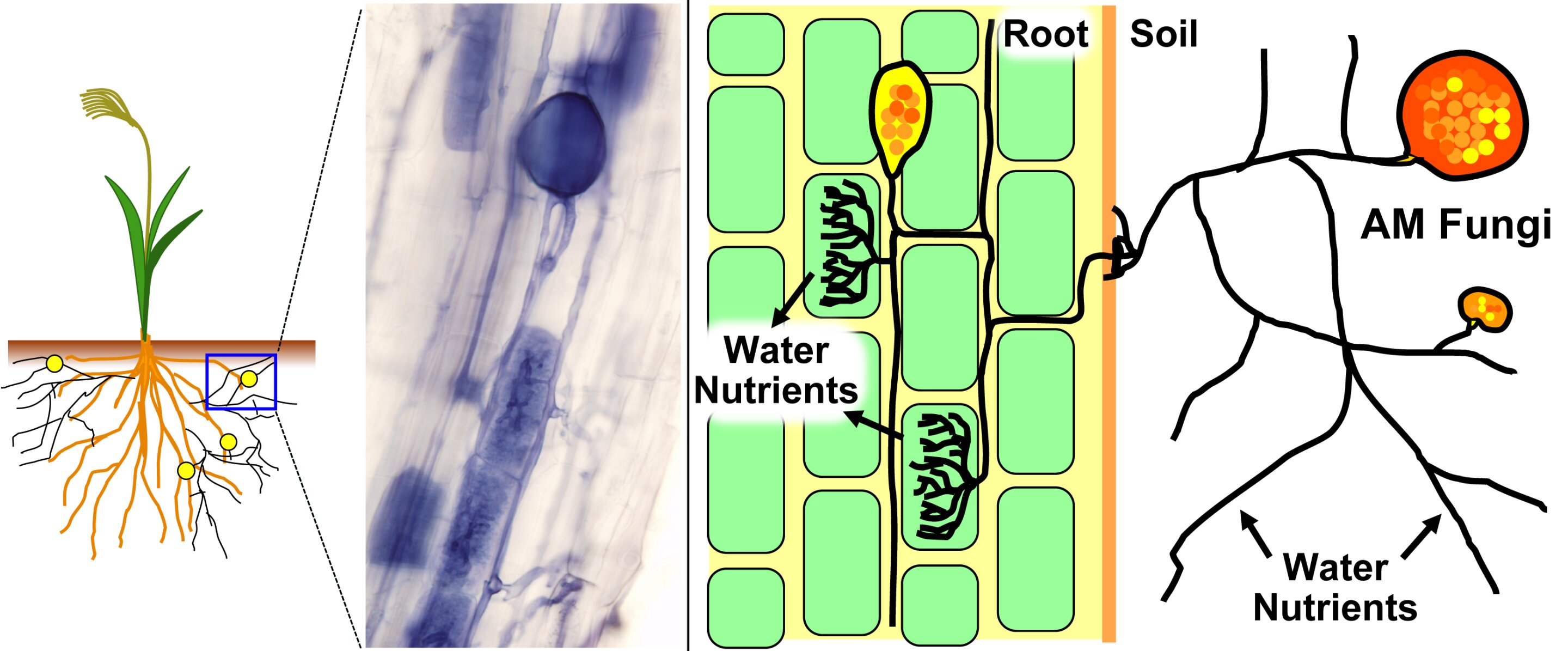
Figure 1: AMF symbiosis with plant roots — arbuscules and vesicles.
AMF are a specialized group of fungi belonging to the phylum Glomeromycota. Unlike many other soil fungi, AMF are obligate symbionts, meaning they cannot complete their life cycle without associating with a host plant. This partnership — known as arbuscular mycorrhiza — occurs in approximately 80% of all land plant species (Smith & Read, 2008).
In this mutualistic relationship, the plant provides the fungus with carbohydrates derived from photosynthesis. In return, AMF's extensive network of fine thread-like hyphae extends far beyond the plant's root system into the soil — accessing tiny pores unavailable to the plant alone, dramatically increasing uptake of phosphorus (P), nitrogen (N), zinc (Zn), and copper (Cu) (Marschner & Rengel, 2012).
Key Structural Organs of AMF
🌿Arbuscules: Highly branched, tree-like structures formed inside plant root cells. These are the primary sites for nutrient exchange between fungus and plant — ephemeral, lasting only a few days before reabsorption.
💧Vesicles: Lipid-filled storage organs formed by the fungus within or between root cells. They serve as energy reserves and can act as propagules for new fungal growth.
🔗Intraradical hyphae: Fungal hyphae growing within the root cortex, connecting arbuscules and vesicles throughout the root tissue.
🌐Extraradical hyphae: Hyphae extending from the root surface into the surrounding soil — forming the vast network responsible for nutrient acquisition across a much larger soil volume.
Methodology
Extracting & Staining AMF from Roots
To study AMF colonization, researchers must first make the fungal structures visible within the plant roots. This involves a two-step process: clearing and staining.
Step 1 — Root Clearing
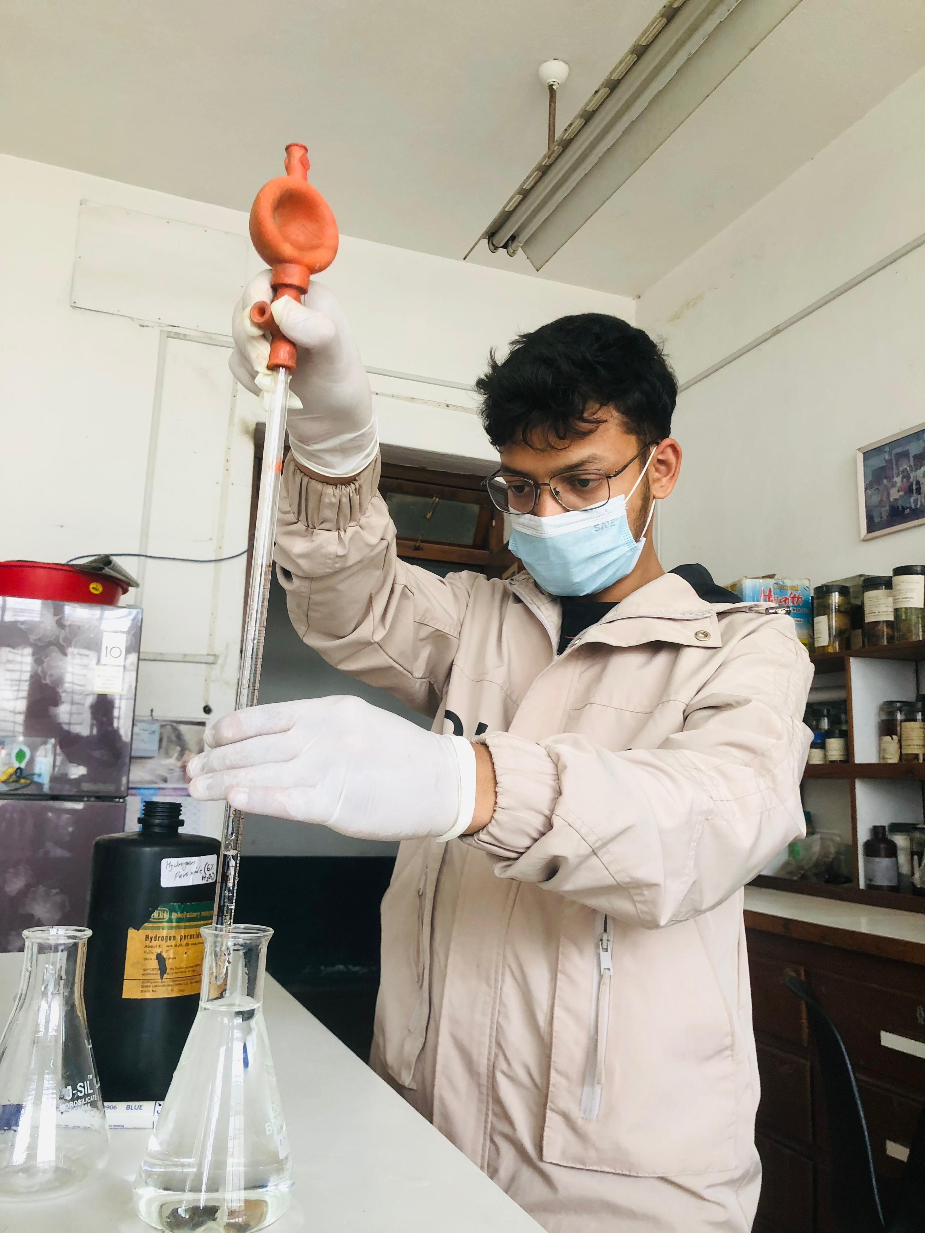
Figure 2: Preparing chemicals for the root staining process.
The purpose of clearing is to remove the plant's cellular contents, pigments, and other compounds that would otherwise obscure the fungal structures — making root tissue transparent so stained fungi can be clearly observed.
→Procedure: Fresh root samples are washed to remove soil, then immersed in 10% KOH solution and incubated at 90°C for several hours. This alkaline treatment dissolves cytoplasm and organic matter, leaving the fungal chitinous cell walls intact (Phillips & Hayman, 1970).
→Rinsing: After KOH treatment, roots are thoroughly rinsed with distilled water to remove residual alkali before staining.
Step 2 — Root Staining
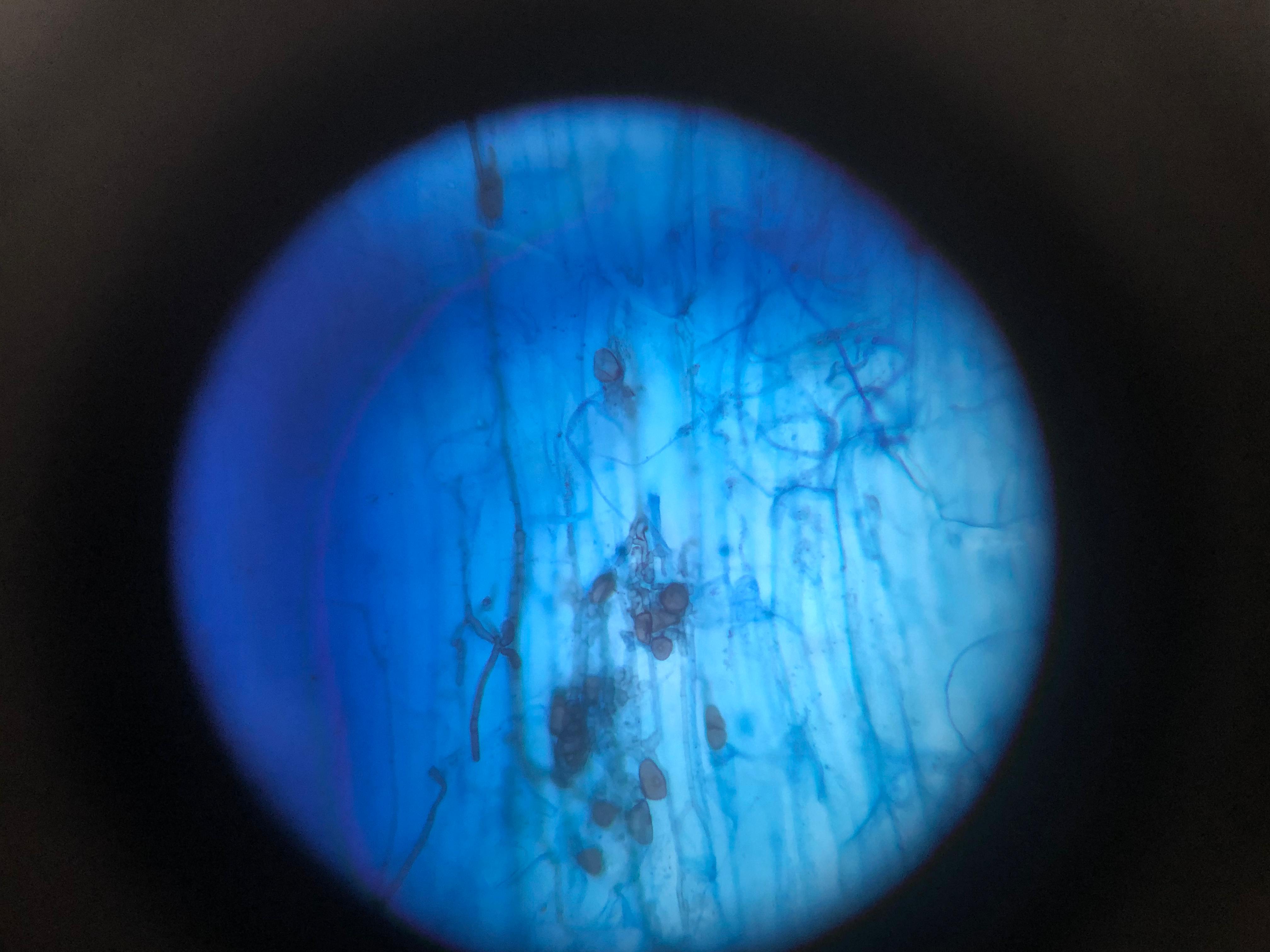
Figure 3: Microscopic view of AMF-colonized roots (my research).
Once cleared, the fungal structures are still largely transparent. Staining makes them visible by selectively binding to components of the fungal cell walls.
→Acidification: Roots are soaked in 1% HCl to neutralize residual KOH and enhance stain uptake by fungal structures.
→Staining Solution: The most commonly used stain is Trypan Blue (0.05–0.1% in lactic acid-glycerol-water). Roots are incubated at 60–90°C for 10–30 minutes. The stain selectively binds to chitin in fungal cell walls, turning hyphae, arbuscules, and vesicles a distinct blue-black (Koske & Gemma, 1989).
→Destaining: Roots are transferred to acidified glycerol to remove excess stain from plant tissue, enhancing contrast between clear plant cells and stained fungal structures.
Quantification
The Gridline Intersect Method
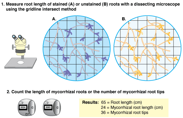
Figure 4: Gridline intersection for calculating colonization %.
Once AMF structures are stained and visible, the next step is to quantify the level of root colonization. The gridline intersect method is a widely adopted, efficient, and statistically robust technique for this (Newman, 1966).
1Root Preparation: Stained root segments are arranged on a microscope slide and mounted in preserving solution under a coverslip.
2Microscope Setup: The slide is placed under a compound microscope fitted with an ocular grid (etched reticle or digital overlay). Magnification is typically 100–200×.
3Systematic Scanning: The observer scans the entire length of root segments using a serpentine stage movement, ensuring complete coverage.
4Counting Intersections: Each time a gridline intersects a root segment, this is counted as one observation point. The total count represents the overall sample.
5Assessing Colonization: At each intersection, the observer determines if any AMF structure (hyphae, arbuscule, or vesicle) is present. If so, that intersection is recorded as "colonized."
6Data Recording: Total intersections and colonized intersections are both recorded for each sample.
7Calculation: Percentage colonization is computed using the formula below.
Colonization Formula (Newman, 1966)
% Colonization
=Number of Colonized IntersectionsTotal Number of Root Intersections×100
This method provides a reliable estimate of the proportion of the root system that AMF actively colonizes. It is particularly useful for comparing colonization levels across different plant species, soil types, environmental conditions, or experimental treatments.
Broader Impact
The Broader Implications of AMF Research
The ability to accurately extract, stain, and quantify AMF colonization is foundational for advancing our understanding of these critical symbionts. Research utilizing these methods helps us to:
🌱Optimize Agricultural Practices: Develop sustainable farming methods that enhance natural AMF populations, reducing the need for synthetic fertilizers and pesticides — directly relevant to Nepal's farming communities.
💪Improve Plant Resilience: Breed and select plant varieties forming stronger AMF associations, increasing tolerance to drought, salinity, and disease (Auge, 2001).
🌍Facilitate Ecological Restoration: Utilize AMF inoculants to aid in revegetation of degraded lands and promote biodiversity recovery in damaged ecosystems.
🔬Understand Soil Health: Gain deeper insights into complex interactions within the soil microbiome and their impact on nutrient cycling and soil structure — foundational to Krishi Sarathi's research mission.
🇳🇵 Relevance to Nepal
This research is directly applied in Krishi Sarathi's ongoing work — including Anup Pudasaini's thesis on the Effect of AMF and Phosphorus on Soil Nutrient Availability for Growth and Yield of Potato, funded by Nepal's Ministry of Forest and Environment and NAST under the Ecosystem-Based Adaptation framework.
Academic References
Sources & Citations
Auge, R. M. (2001). Water relations, drought and VA mycorrhizal symbiosis. Mycorrhiza, 11(1), 3–42.
Koske, R. E., & Gemma, J. (1989). A modified procedure for staining roots to detect mycorrhizas. Mycological Research, 92(4), 486–490.
Marschner, P., & Rengel, Z. (Eds.). (2012). Marschner's Mineral Nutrition of Higher Plants (3rd ed.). Academic Press.
Newman, E. I. (1966). A method of estimating the total length of root in a sample. Journal of Applied Ecology, 3(1), 139–145.
Phillips, J. M., & Hayman, D. S. (1970). Improved procedures for clearing roots and staining parasitic and vesicular-arbuscular mycorrhizal fungi. Transactions of the British Mycological Society, 55(1), 158–161.
Smith, S. E., & Read, D. J. (2008). Mycorrhizal Symbiosis (3rd ed.). Academic Press.
More Research
Explore Our Other Research
AMF is just one piece of Krishi Sarathi's research into sustainable soil management. Discover our work on Biochar technology and how both innovations are transforming Nepali agriculture.